

An EKG, the electrocardiogram is the friendly name of a device that is used often by medical professionals and that is a suitable tool to analyze the electricity of the heart. The device explores the nature of the disease at a molecular and genetic level, highlighting the effective management of numerous cardiovascular conditions.
This extensive article will dig up to the core of EKG, analyzing its significance, the basic principles of its operations, and the situations when such a test may be recommended. Whether you work as a healthcare provider and you seek to bring your knowledge up to par or you are a patient who is making efforts to go through the intricacies of cardiovascular disease, the following article seeks to give you the essential information you need for you to appreciate the value of EKGs in the contemporary medical practices today.
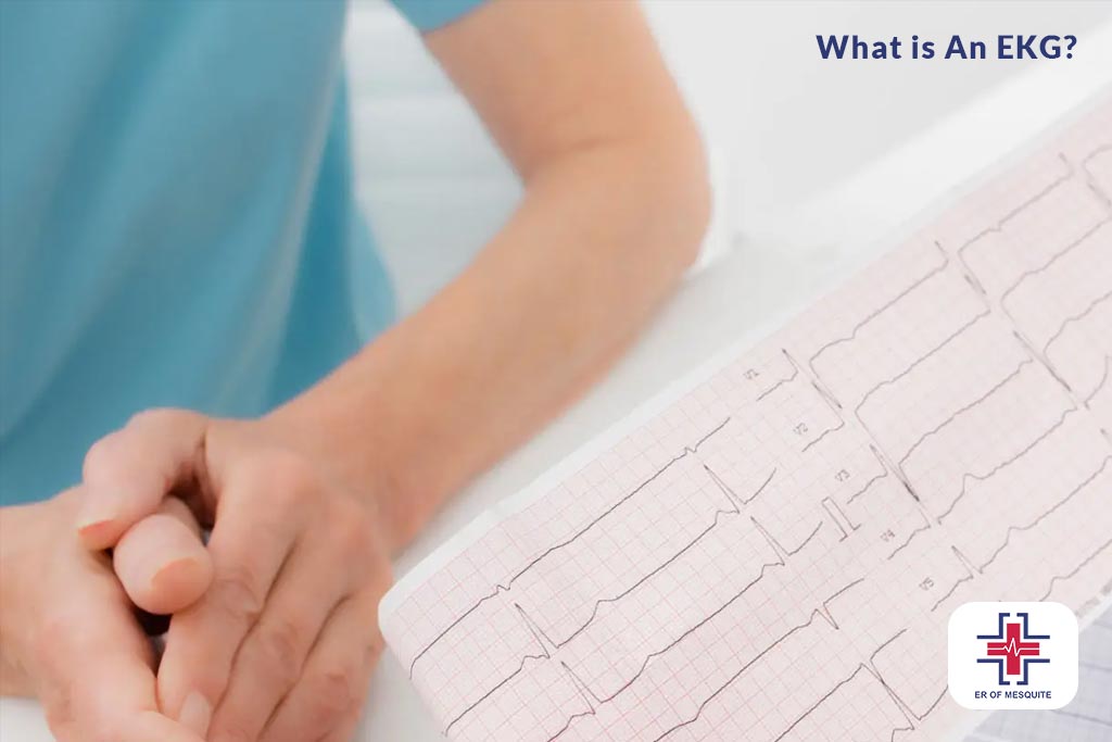
What is An EKG?
An electrocardiogram is a kind of medical test that records the electrical activity of the heart during a period and uses electrodes placed on the skin to do so. These electrodes then capture the subtle skin’s electrical changes that are generated by the heart muscle as the latter electrophysiologically depolarizes and repolarizes constantly while the GS heartbeat beats. It is a widely loved and painless procedure utilized to rapidly shed light on cardiovascular diseases and maintain tracking of the heart’s well-being in a variety of situations.
EKGs are used to:
- Assess the heart’s rhythm.
- Detect global cardiac muscle blood restriction caused by atherosclerosis.
- Diagnose a heart attack.
- Evaluates the dysrhythmia and other forms of abnormalities of the heart, like ventricular hypertrophy.
- Keep tracking heart disease risk occurrence.
An EKG provides two major kinds of information: the brief (time interval) cognitive process required for electrical waves to navigate the muscle meat of the heart, showing slow, fast, or irregular light dissipation and the exact amount of electric activity passed through the heart muscle (to indicate the size of as well as over-worked regions).
What Are the Risks and Drawbacks?
The EKG analysis is one of the commonly used tests in medicine as it is very useful in checking numerous types of heart diseases. The test kits can be found in any developed medical facility and are relatively safe, accurate, affordable, and user-friendly to administer.
Possible risks of EKG include:
- Discomfort or allergic reaction caused by the device which is stuck to your arms and chesty with the help of single-use adhesive electrodes made of agrochemicals
- The electrode’s constant contact with the tissue tends to produce tissue breakdown associated with the persistence of the signal throughout the outdoor ambulatory ECG.
- The feelings of lightheadedness or fainting that are associated with heart problems, including a heart attack, are examples of such events.
While an EKG gives us valuable data to help detect heart problems, it has a few limitations as well. Among them:
- An EKG taken in the resting position cannot reveal issues that take place while tracing the tracking signal and some of these issues like tachycardia, are occasional. An ambulatory EKG may be required after a resting EKG to assess a suspected problem if it has not been able to do so.
- An EKG may show an NAF (normal or near-to-normal) reading with diverse types of heart diseases, including CAD (coronary artery disease). For others the doctors will order different tests, namely angiography, to reveal the problems.
- Electrocardiogram may be an attending feature of the disease but it’s unlikely to be the sole reason. Electrocardiograms(EKG) bring into light small imperfections that eventually prove to be of insignificant medical relevance after careful examination.
Types of EKG
Different types of the EKGs can be used. Their choice is determined by the clinical diagnostic situation and by the information they will elicit. Here are the main types of EKGs:
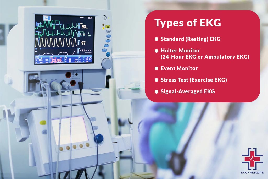
Standard (Resting) EKG:
- It is the main one and is the one used mostly in doctor’s offices or hospitals.
- A patient lies on a bed – electrodes are attached to the arms, legs, and chest.
- The EKG can interpret the heart’s electrical waves for a short time, typically about 10 seconds.
- It is used mostly for heart problems such as arrhythmias or a past heart attack that do not necessarily require remedial actions in a resting state.
Holter Monitor (24-Hour EKG or Ambulatory EKG):
- In this kind of procedure, you are required to put on portable EKG equipment for 24 hours or longer, without this being intrusive to your daily routines.
- It records continuous information related to the heart’s electrical activity and is used for detecting the following conditions that rarely manifest on a standard EKG: irregular rhythm (arrhythmia).
Event Monitor:
- The same integral principle applies to this device also, as it is carried by a person almost 24 hours a day for some weeks.
- It focuses only on the electrical pattern produced by the heart in particular periods. It will either record them automatically when the pulse produces abnormal rhythms or when the patient activates it when he notices the symptoms.
- This type is extremely beneficial for detecting and restoring abnormalities that occur with rare frequency.
Stress Test (Exercise EKG):
- A stress Test is carried out during the patient’s exercise on a treadmill or stationary bike (while the heart works hard and beats fast) to check if it is under stress as well.
- This test is of vital importance as it provides insights into the amount that the heart can handle, which in turn is an indication of the presence or lack of coronary artery disease or the level of a person’s cardiac treatment.
Signal-Averaged EKG:
- An EKG adds a degree of detail to the classic isophase and it involves averaging multiple readings over time to make a more comprehensive and accurate depiction of the heart’s electrical activity.
Different types of EKGs were adapted to target specific diagnostic needs, ensuring the delivery of crucial information to confirm diagnosis and treat various heart conditions.
What Are 3 Reasons a Person Would Get An EKG?
EKG in cardiology is a crucial and basic diagnostic tool that is used by many people in the field of cardiology. It reveals the electrical activity of the heart and becomes a valuable device in many clinical circumstances for appraisement of heart conditions. An EKG (also called an electrocardiogram) is often performed to shed light on heart disorders and is popularly used within the scope of medical situations. This lies in the depth of the critical aspects of the EKG method which in turn explains why a patient can be submitted to an EKG examination.

Symptom Evaluation
The EKG is the reason for the first visit because it is suitable to clarify if some symptoms are possibly indicative of having a heart condition. Often the chest pain becomes exacerbated, presented with dyspnea either shortness of breath or lightheadedness, with fast heart rate and palpitation in a situation where EKGs are done to establish any relationship with the issue and the symptoms. Possibly, these kinds of bouts could demonstrate to you that some heart problems might occur, such as arrhythmias, coronary artery disease, or previous heart attack. EKG is responsible for one particular job which is to make the problem very clear to the eye, where the heart is closing the deal, and also detects any abnormal electrophysiologic activity.
Routine Health Screening
Electrocardiograms done during check-ups are routinely recommended, particularly for patients who have a previous history of cardiovascular disease or are genomically predisposed to acquiring such. These are high systolic blood pressure, high cholesterol, diabetes, a family history of cardiovascular disease, or smoking patients should have to be they. In this line, the test is also recommended carried out among aged people so that there would be follow-up in any progress in the health status of people. An important way is doing EKG screening constantly which makes it possible to detect emerging heart abnormalities earlier than any other way and intervention can be made if the downward spiral starts. And so, a person suffering from the disease can feel healthy.
Pre-Operative Assessment
The physiological condition of the heart is assessed carefully before any major surgery as the cardiovascular system may undergo life-threatening stress situations from the surgery. It can be viewed as taking place by measuring the heart’s electric activity that in essence will give the complete picture of the heart’s performance. This is critical, especially for those prior patients who had a recorded history of heart failure or possess the major risk factors of heart disease. An EKG is needed to ascertain the health of the cardiorespiratory system to determine and select appropriate anesthesia for safety and then also plan for unanticipated cardiac adverse effects in the future.
How to Read EKG Strips?
It is one of the vital devices used in the diagnosis of a vast spectrum of cardiovascular ailments, from timid rhythm abnormalities to life-threatening heart attacks. EKG strip reading can be considered the most fundamental skill gained for healthcare professionals, but also useful to anyone interested in deeper knowledge about cardiac web fundamentals. This post serves as a useful introduction to knowledge of typical EKG strip interpretation.

Understanding the Basics
A strip chart is a graphical display of the heart’s electoral data, which captures all electrical activity for a given period and prints the information on graph paper. The axis of the EKG paper which faces the horizontal direction (time) ranges from 1 to 4 in height. The paper is marked with a grid of small and large squares: small squares, each 0.04 seconds in length, and large, 5 x 5 squares of small squares, 0.20 seconds in length, each representing one simplicity.
The Components of an EKG Strip
- P Wave: The first wave lighted on the EKG strip, which in order they happen in the body are: the atrial depolarization, or we can call it, contraction of atria. A usual P wave is a smooth, rounded affair that is not greater than 0.11 seconds in duration.
- QRS Complex: The P wave as seen after it, the QRS complex, is the section which is the most apparent and is the symbol of bipolar intracardiac depolarization, that is, the contraction of the ventricles. NTC most often lasts from 0.6 to 0.10 microseconds. The QRS complex is the indicator that is useful for determining the ventricular rate and rhythm.
- T Wave: The T wave slowly turns up when the QRS complex has ended and is ventricular repolarization, the time when ventricles are prepared for the next beat. A familiar T wave is finished after the QRS complex and points in the same direction as the QRS complex.
- PR Interval: This time is formed by the period starting from P wave delivery to QRS complex beginning and shows how long that impulse needs to run from atria to ventricles. The regular PR interval will be anywhere between 0.12 to 0.20 seconds.
- ST Segment: The P wave in the middle is consistent with the QRS complex followed by the T wave. Heart muscle relaxation (represented by J-point) in comparison with the baseline (isoelectric line) may signal myocardial ischemia or infarction.
Step-by-Step Analysis
- Determine the Heart Rate: QRS complexes number for a six-second EKG strip should be counted and then multiplied by 10 to get the heart rate. This will provide you with beats per minute and is the parameter you should adjust first.
- Assess the Rhythm: Make sure the rhythm is regular by calculating the space between R-waves and taking an R-R interval. If the intervals are the same, therefore, rhythm is periodic, and otherwise rhythm is interrupted.
- Analyze the Waves and Segments: Evaluate the P waves, QRS complexes, T waves, PR intervals as well as ST segments to diagnose any abnormalities in size, form, and timeframe.
- Interpret the Findings: Combine the data from your investigation to get conduction of a cardiac rhythm together. Such as the P waves which are not present and QRS complexes which are irregular, these can indicate atrial fibrillation.
Reading an EKG strip appears difficult initially, yet with repeated exercise, it would represent a helpful skill to use for diagnosis as well as monitoring of overall heart health. Cardiography based on EKG strip interpretation is an important skill for healthcare learners, current practitioners, or popular audiences. It can reveal more information about cardiac function, and make the public health situation better
What Does a Normal EKG Look Like?
A usual EKG (electrocardiogram) just looks like a consistent rhythmic series of waves and complete complexes in each heartbeat. The initial movement on each EKG strip is the P wave, a small upward curve of which indicates the atrial, upper chambers of the heart electric impulse that prompts the atria to contract. We have this right after them, a complex QRS which is somehow the most striking part of the tracing. The QRS complex which has the shape of a rapid rising step stands for rapid depolarization of the heart’s ventricles, which are the bottom chambers of the heart.
The T wave (a smaller upward pulse) that follows the QRS complex represents the ventricular repolarization period (the interval between two successive beats) when the ventricles are preparing for the next beat. This wave is calmer and wider than the rectangle QRS complex or rectangle in general.
The Section between the P wave and the QRS Complex is abbreviated as the PR interval. It shows the interval of time when the electrical impulse transmission occurs from the A-V node to the A-V bundle. PR is a crucial interval in the study of PR because it represents the time during which the signal is going through the AV node, the cardiac electrical junction between the right and the left atrial.
Moreover, another important designation is the area connecting the QRS complex to the T wave, known as the ST segment. No changes and no deviation from the baseline should be visible on this segment to prove the normal conduction and repolarization.
What Does an Abnormal EKG Look Like?
An abnormal EKG can show different features that suggest a possible cardiac problem and these are hidden in different alterations of the known patterns of the EKG in the case of a healthy heart rhythm.
An abnormal EKG can be signified by an arrhythmias of the electrocardiograph. In this mismatch, R is the R-R intervals variable which is observed as heartbeats that are not evenly spaced such as in atrial fibrillation and the irregularity in the baseline is because of the activities of atria that are irregular.
Changing patterns as well should be looked upon as indications of problems. The dynamic waveform is a hallmark of malfunctioning. For example, grossly enlarged P waves or those with an abnormal development may indicate an atrial enlargement, while changes in QRS complex form, such as widening (over 120 milliseconds), may suggest a delay in the electrical conduction through the ventricles, a condition observed in bundle branch block. Likewise, T Wave Inversions can also be a sign of ischemia or other cardiac issues and U waves may mean Hypokalemia.
Rhythm or rate shifts and changes in the intervals of the relevant features are also helpful in determining the diagnosis through an EKG. A PR interval extended for a long time could likely be due to a first-degree heart block, which features an arrhythmia in the AV node causing the electrical impulse to travel slowly. However, an intervention with a short PR interval could take place in anomalies such as the Wolf-Parkinson Infrared syndrome, which has the electrical impulse bypass the normal way. An ST segment change, especially. when the amplitude is increased, may indicate an acute myocardial infarction, while the decrease suggests ischemia.
Finally, the distortions of irregularities in the natural zero-to-tachycardia rhythm, also known as bradycardia, can also significantly affect the EKG. The proximity of QRS waves in tachycardia is conspicuous and the distance between them is widely separated in bradycardia.
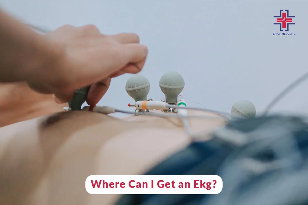
Where Can I Get an EKG?
They are done in healthcare institutions, including urgent care, and ED, among others, depending on the importance and details of the health concern. Here are the typical places where an EKG can be performed:
- Doctor’s Offices: Some primary care physicians and cardiologists may be able to execute an EKG (electrocardiogram ) in their offices as part of regular exams as well as if there is such a suspicion of – heart disorder.
- Hospitals: Amidst many activities happening in hospitals, they are the heart of emergency rooms where instant assessment of patients’ heart function is the common need.
- Urgent Care Centers: When the urgency is not to declare, a non-life-threatening heart condition, EKG testing is offered, and prompt attention is not necessary.
- Diagnostic Labs and Clinics: These dedicated diagnostic centers and clinics will also do EKGs such as upon the recommendation of a medical practitioner.
Conclusion:
In the final analysis, the EKG is an indispensable piece of equipment used in cardiology to give details about the electrical activity of the heart and the general health of the patient. This non-invasive simple test can identify multiple cardiovascular disorders, such as electronic instability (arrhythmias), artery narrowing (ischemia), muscular infractions (myocardial infarction), and other structural faults. Learning how and what type of EKG to apply is indeed very important. Therefore, you need to be aware of whether you are experiencing symptoms, need a routine check due to the existing risk factors, or perform surgery preparation for this EKG.
Such symptoms could be an alarm for requiring an EKG or just need the review of the cardiovascular system, so you might consider going to the city’s ER. The facility offers a full range of technologically advanced diagnostic components and is staffed with experienced medical doctors who can carry out thorough cardiac evaluations, including EKG testing. If you are looking for a reliable EKG test you should visit Walk in Clinic Mesquite immediately.
FAQ
How far back can an EKG detect a heart attack?
One of the main features of an EKG is the ability to identify marks of heart attacks forever since the ECG compares the results of the present heart rate to the normal ones that the doctor has already compiled before. The changes in the heart muscle from old attacks can leave the permanent markers on an EKG – as well as Q waves.
What does an EKG show?
An EKG provides the electrical activity recorded of the heart muscle. It is capable of showing us our heart rate, irregularities, and rhythm, which helps doctors diagnose conditions such as arrhythmias, heart attacks, and other cardiovascular disorders.
What does EKG stand for?
EKG denotes an Electrocardiogram, which is listed alongside ‘Elektrokardiogramm’, the term in German. It is known as Electrocardiogram in the common terminology, and it is also referred to as ECG.
How much does an EKG cost?
The price of EKG is almost too generic and depends on a lot of parameters such as location and facility and can range from somewhere between $50 to $300 when there is no insurance. Some health penalties might provide some partial to full coverage of foregoing EKG expenses.