Your doctor mentions ECG and CT scan in the same breath. Panic sets in. Are they interchangeable? Is one more serious than the other?
These two powerful diagnostic tools offer unique insights into your heart’s health, but they’re far from identical. Understanding their differences could be the key to unlocking crucial insights about your cardiovascular well-being.
From detecting irregular heartbeats to unveiling hidden arterial blockages, ECGs and CT scans play distinct roles in cardiac care. But when do you need which?
What is an ECG?
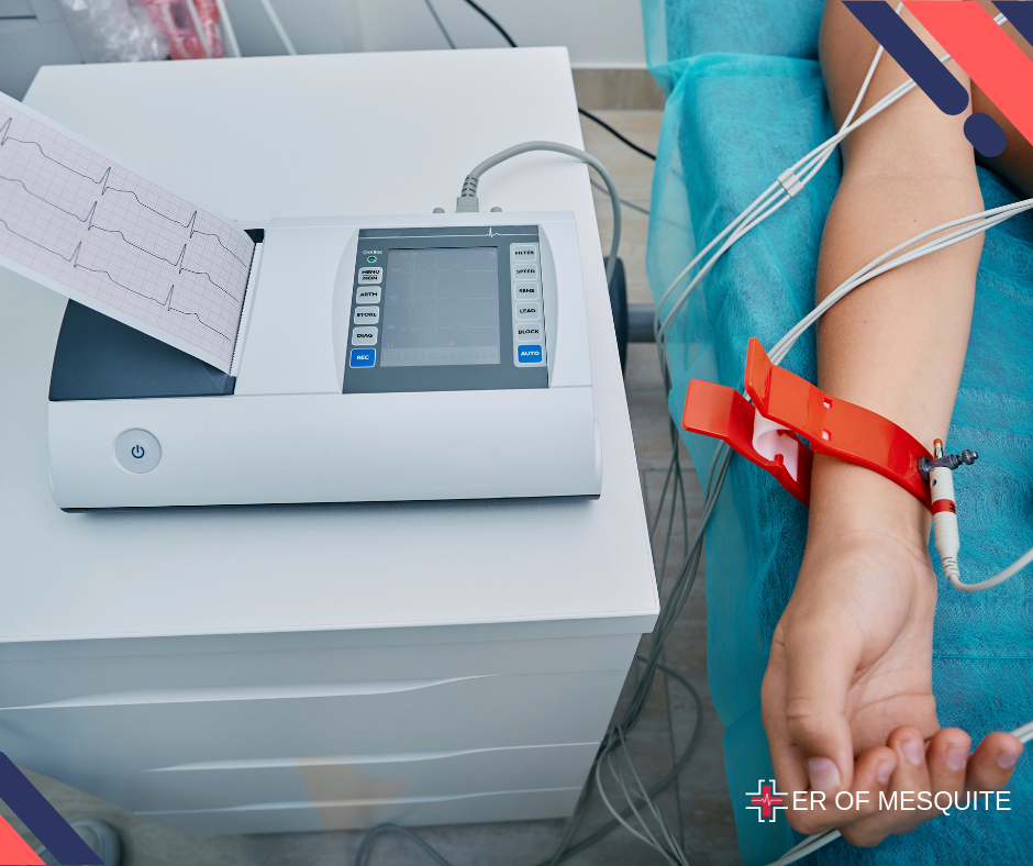
An electrocardiogram (ECG or EKG) is a non-invasive test that measures the heart’s electrical activity. It’s typically the first test done when a patient shows symptoms like chest pain, shortness of breath, or palpitations.
The ECG records the timing and strength of these electrical signals as they move through the heart, displaying them as waves on a monitor or printed on paper.
ECG Uses
- Detecting Arrhythmias: It identifies irregular heart rhythms that indicate conditions such as atrial fibrillation or ventricular tachycardia.
- Diagnosing Heart Attacks: The ECG shows changes in the heart’s electrical patterns that suggest a heart attack or myocardial infarction.
- Assessing Heart Damage: It provides information about past heart attacks or ongoing damage to the heart muscle.
- Monitoring Heart Health: It is useful for routine check-ups, especially for patients with a history of heart disease.
ECG Types
The three main types of ECG are:
- Resting ECG: During a resting ECG, you lie down without moving to prevent interference from muscle activity. This test typically lasts between five to 10 minutes.
- Ambulatory ECG: With an ambulatory ECG, you wear a portable device for at least 24 hours, allowing normal movement. It is used to capture intermittent symptoms that may not appear during a resting ECG, particularly useful after a heart attack. You may record symptoms to compare with the ECG.
- Exercise Stress Test: This ECG assesses heart function during physical activity, such as riding a stationary bike or walking on a treadmill. It takes about 15 to 30 minutes and may involve the use of medication to evaluate heart response.
Learn How ECGs Prevent Heart Diseases
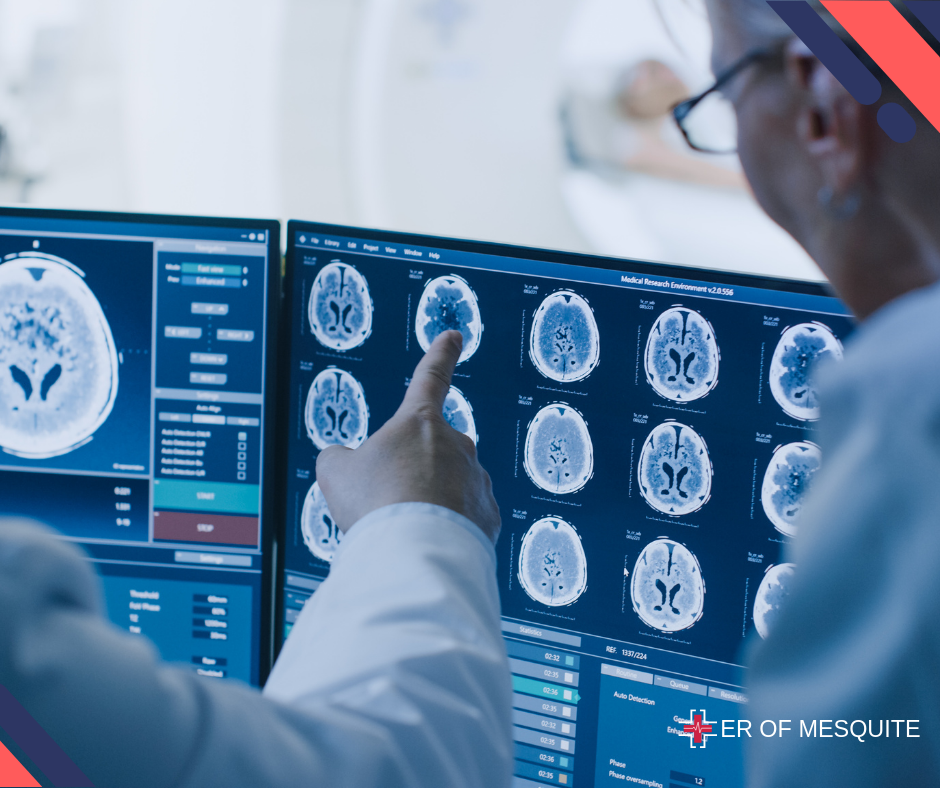
What is a CT Scan?
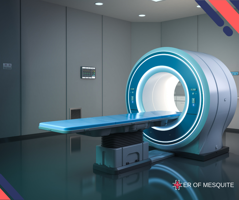
A CT scan, also known as computed tomography, is an advanced imaging test that utilizes X-rays and computer processing to create detailed cross-sectional images of the body.
When examining the heart, a specialized type called cardiac CT or coronary CT angiography is employed. This test offers detailed views of the heart’s anatomy, including the coronary arteries, aiding in the assessment of structural issues and blockages.
CT Scan Uses
- Detecting Coronary Artery Disease (CAD): Identifies blockages or narrowing in the coronary arteries that can cause chest pain and heart attacks.
- Assessing Heart Anatomy: Provides clear images of the heart’s valves, chambers, and overall structure, assisting in the diagnosis of congenital heart defects or other structural issues.
- Evaluating Aortic Diseases: Helps in diagnosing conditions like aortic aneurysms or dissections.
- Planning Treatment: Essential for planning interventions such as stenting or surgery by providing detailed maps of the heart’s internal structures.
CT Scan Types
- Cardiac CT Angiography (CTA): This CT scan is focused on capturing images of the coronary arteries to detect blockages or narrowing that may lead to conditions such as coronary artery disease (CAD). It provides detailed images showing how blood flows through the heart.
- Coronary Calcium Scoring (CAC): This CT scan measures the amount of calcium deposits in the coronary arteries. These deposits can indicate the presence of atherosclerosis (plaque buildup) and help assess the risk of heart disease.
- Cardiac CT for Structural Imaging: This type of CT scan provides detailed images of the heart’s structure, including its chambers, valves, and overall anatomy. It aids in diagnosing congenital heart defects and other structural abnormalities.
- CT for Aortic Imaging: CT scans can also be used to examine the aorta, identifying conditions such as aortic aneurysms (abnormal bulges in the aortic wall) or aortic dissections (tears in the aortic wall).
ECG vs CT Scan
Purpose
- ECG: Focuses on monitoring the heart’s electrical activity, aiding in diagnosing arrhythmias, heart attacks, and overall heart function assessment.
- CT Scan: Provides detailed anatomical images that assist in diagnosing structural heart diseases, coronary artery disease, and planning treatment strategies.
Procedure
- ECG: Quick and non-invasive; involves placing electrodes on the skin to record heart activity.
- CT Scan: The CT scan procedure is more involved, typically requiring the injection of contrast dye and taking longer to complete.
Information Provided
- ECG: Displays the heart’s electrical patterns and rhythms, helping to identify abnormalities.
- CT Scan: Produces detailed images showing the heart’s structure and the condition of coronary arteries.
Use in Diagnosis
- ECG: Primary diagnostic tool for evaluating symptoms such as chest pain, palpitations, and suspected heart attacks.
- CT Scan: Used as a follow-up or supplementary test when investigating suspected structural issues or coronary artery disease.
When Do You Need an ECG?
You may require an ECG if you have the following symptoms:
- Chest pain
- Palpitations
- Dizziness or fainting
- Shortness of breath
- Fatigue
Routine ECGs are also recommended if you have risk factors for heart disease, such as high blood pressure, high cholesterol, diabetes, or a family history of heart disease.
When Do You Need a CT Scan?
A CT scan may be recommended if:
- You have unexplained chest pain and your doctor suspects coronary artery disease.
- There is a need to evaluate the structure of your heart and blood vessels.
- Planning for surgical procedures or interventions is required.
- There is a need to diagnose or monitor aortic diseases or other structural abnormalities.
Key Takeaway
Both ECGs and CT scans play crucial roles in diagnosing and managing heart disease, each with its unique advantages. An ECG swiftly provides essential information about the heart’s electrical activity, making it invaluable for diagnosing arrhythmias and detecting heart attacks.
In contrast, a CT scan offers detailed anatomical images, vital for identifying structural heart problems and planning effective treatments.
At ER of Mesquite, we specialize in delivering precise diagnostic results and comprehensive cardiac care. Whether you need an ECG for immediate assessment or a CT scan for detailed evaluation, our team of expert radiologists is committed to prioritizing your heart health.
Schedule Your Heart Health Consultation
FAQs
What is an ECG and CT scan?
An ECG (electrocardiogram) measures the electrical activity of the heart to detect arrhythmias and heart attacks. A CT scan (computed tomography) uses X-rays to create detailed images of the heart’s structures, aiding in diagnosing structural issues and planning treatments.
Does a CT scan show heart problems?
CT scan can reveal heart problems by providing detailed images of the heart’s structures, including blockages in the coronary arteries. It is particularly useful for diagnosing coronary artery disease and other structural heart issues.
What ECG Cannot detect?
An ECG cannot detect structural heart abnormalities, blockages in the coronary arteries, or issues with the heart valves. It primarily measures electrical activity and rhythm, not anatomical features.